Epilepsy & Seizures
Protocadherin19 clustering epilepsy
Mar. 06, 2024
MedLink®, LLC
3525 Del Mar Heights Rd, Ste 304
San Diego, CA 92130-2122
Toll Free (U.S. + Canada): 800-452-2400
US Number: +1-619-640-4660
Support: service@medlink.com
Editor: editor@medlink.com
ISSN: 2831-9125
Toll Free (U.S. + Canada): 800-452-2400
US Number: +1-619-640-4660
Support: service@medlink.com
Editor: editor@medlink.com
ISSN: 2831-9125
Nearly 3,000 illustrations, including video clips of neurologic disorders.
Every article is reviewed by our esteemed Editorial Board for accuracy and currency.
Full spectrum of neurology in 1,200 comprehensive articles.
Listen to MedLink on the go with Audio versions of each article.
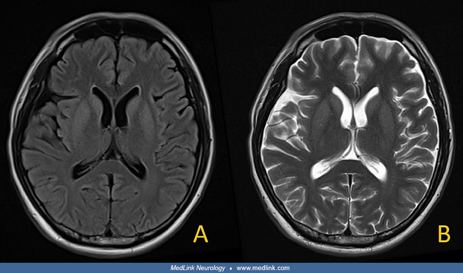
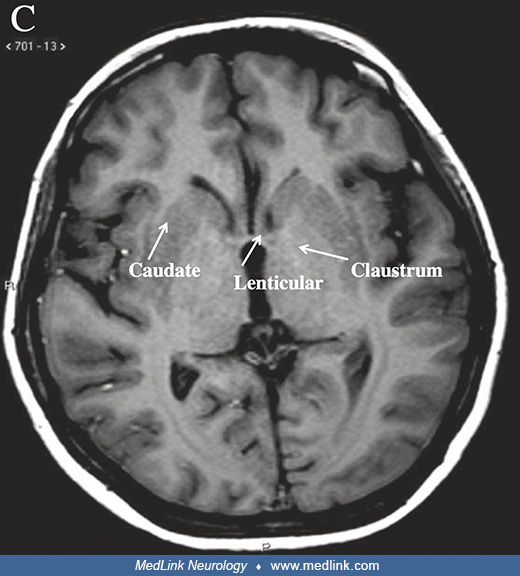
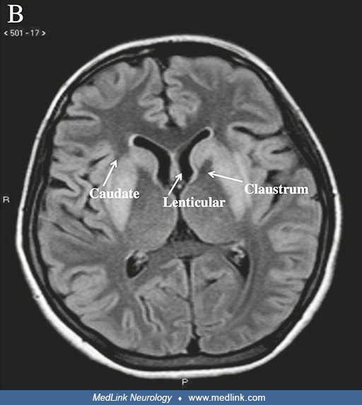
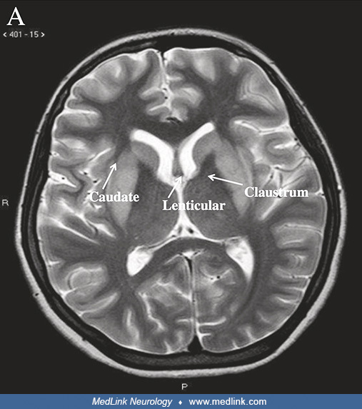
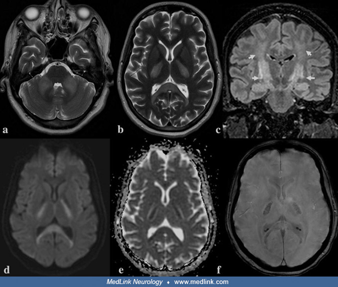
T2-weighted MRI in a 41-year-old woman who presented with acute parkinsonism following slow correction of hyponatremia resulting from repeated vomiting. MRI showed symmetrical hyperintensities in caudate nuclei (small, thin, black arrow) and putamena (long black arrow), with sparing of the globi pallidi (broad black arrow), consistent with extrapontine myelinolysis. (Source: Sajith J, Ditchfield A, Katifi HA. Extrapontine myelinolysis presenting as acute parkinsonism. BMC Neurol 2006;6:33. Creative Commons Attribution license [CC BY], https://creativecommons.org/licenses/by/2.0.)