Mevalonate kinase deficiency
Oct. 17, 2023
MedLink®, LLC
3525 Del Mar Heights Rd, Ste 304
San Diego, CA 92130-2122
Toll Free (U.S. + Canada): 800-452-2400
US Number: +1-619-640-4660
Support: service@medlink.com
Editor: editor@medlink.com
ISSN: 2831-9125
Toll Free (U.S. + Canada): 800-452-2400
US Number: +1-619-640-4660
Support: service@medlink.com
Editor: editor@medlink.com
ISSN: 2831-9125
Nearly 3,000 illustrations, including video clips of neurologic disorders.
Every article is reviewed by our esteemed Editorial Board for accuracy and currency.
Full spectrum of neurology in 1,200 comprehensive articles.
Listen to MedLink on the go with Audio versions of each article.
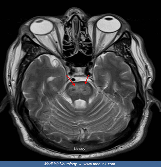
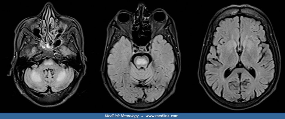
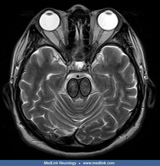
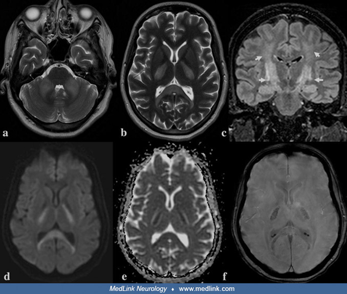
MRI in a 45-year-old alcoholic woman with a recent hospitalization for Wernicke encephalopathy who presented with left hemiparesis and was initially suspected to have had a stroke. (a) Axial T2-weighted image demonstrates high T2-signal intensity in the upper pons, resembling a pig’s snout (the so-called “piglet sign”). (b) The same axial T2-weighted image with the area of edema of the pons is highlighted in green. The transverse pontine fibers (pontocerebellar fibers and median raphe) are most affected and there is sparing of the peripheral (ventrolateral longitudinal) fibers and corticospinal tracts, producing the pig’s snout appearance. The edema involves the right corticospinal tracts (blue arrow), which explains the patient's left-sided hemiparesis. (c and d) DWI and apparent diffusion coefficient (ADC) map corresponding to the restricted diffusion in the pons. (e) Sagittal FLAIR image demonstrates increased FLAIR signal in the pons. (Source: Kusel K, Azzam O, Youssef A, Prentice D. Alcoholic pontine myelinolysis: beware the stroke mimic. BJR Case Rep 2021;7(4):20210005. Creative Commons Attribution license [CC BY], https://creativecommons.org/licenses/by/4.0.)