Neuro-Oncology
Sequelae of treatment of CNS tumors
Jul. 25, 2024
MedLink, LLC
3525 Del Mar Heights Rd, Ste 304
San Diego, CA 92130-2122
Toll Free (U.S. + Canada): 800-452-2400
US Number: +1-619-640-4660
Support: service@medlink.com
Editor: editor@medlink.com
ISSN: 2831-9125
Toll Free (U.S. + Canada): 800-452-2400
US Number: +1-619-640-4660
Support: service@medlink.com
Editor: editor@medlink.com
ISSN: 2831-9125
Nearly 3,000 illustrations, including video clips of neurologic disorders.
Every article is reviewed by our esteemed Editorial Board for accuracy and currency.
Full spectrum of neurology in 1,200 comprehensive articles.
Listen to MedLink on the go with Audio versions of each article.
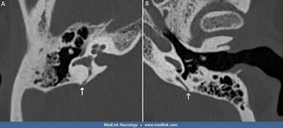
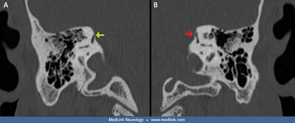
Temporal bone CT scan in a 59-year-old Moroccan man with bilateral posterior and right superior semicircular canal dehiscence. This frontal view shows the dehiscence of the right superior semicircular canal (A) and no bone defect of the left superior semicircular canal (B). (From: Andaloussi TB, Bouqes M, Ouattassi N, et al. Diagnosis and management of bilateral and multiple semicircular canal dehiscence: a case report. Pan Afr Med J 2025;51:12. Creative Commons Attribution 4.0 International [CC BY 4.0] license, creativecommons.org/licenses/by/4.0.)