Epilepsy & Seizures
Pharmacological treatment of epilepsy in neonates
Jan. 31, 2024
MedLink®, LLC
3525 Del Mar Heights Rd, Ste 304
San Diego, CA 92130-2122
Toll Free (U.S. + Canada): 800-452-2400
US Number: +1-619-640-4660
Support: service@medlink.com
Editor: editor@medlink.com
ISSN: 2831-9125
Toll Free (U.S. + Canada): 800-452-2400
US Number: +1-619-640-4660
Support: service@medlink.com
Editor: editor@medlink.com
ISSN: 2831-9125
Nearly 3,000 illustrations, including video clips of neurologic disorders.
Every article is reviewed by our esteemed Editorial Board for accuracy and currency.
Full spectrum of neurology in 1,200 comprehensive articles.
Listen to MedLink on the go with Audio versions of each article.
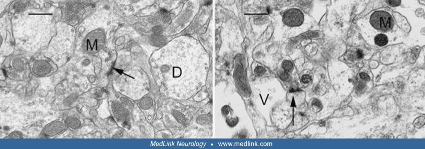
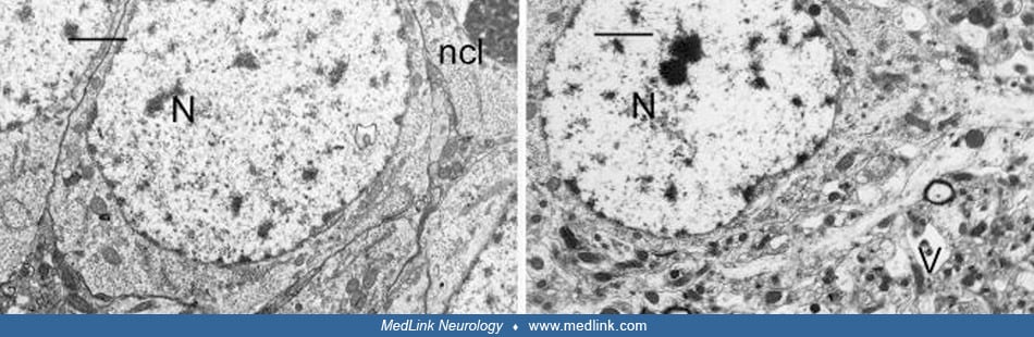
(Left) CA3 region of the hippocampus of a control rat showing a cluster of pyramidal cells with good preservation and integrity of cell membranes and organelles. The nucleus (N) and nucleolus (ncl) are easily identified. A well-preserved electrodense dendritic spine is shown (arrow). Scale Bar = x2.5μm. (Right) CA3 region of the hippocampus of a rat treated by gavage domoic acid for 64 days. Image shows a pyramidal cell with easily identifiable nucleus (N), the cytoplasm and surrounding neuropil with numerous vacuoles (V) of various sizes giving a "Swiss cheese" effect. The neuropil refers to intricate interwoven cell processes including glial processes, synaptic terminals, axons, and dendrites that are interspersed among the nerve cells in the gray matter of the CNS. Scale Bar = x2.5μm. (Source: Pulido 2008. Creative Commons Attribution license. http://creativecommons.org/licenses/by/3.0.)