Epilepsy & Seizures
Epileptic lesions due to malformation of cortical development
Sep. 06, 2023
MedLink®, LLC
3525 Del Mar Heights Rd, Ste 304
San Diego, CA 92130-2122
Toll Free (U.S. + Canada): 800-452-2400
US Number: +1-619-640-4660
Support: service@medlink.com
Editor: editor@medlink.com
ISSN: 2831-9125
Toll Free (U.S. + Canada): 800-452-2400
US Number: +1-619-640-4660
Support: service@medlink.com
Editor: editor@medlink.com
ISSN: 2831-9125
Nearly 3,000 illustrations, including video clips of neurologic disorders.
Every article is reviewed by our esteemed Editorial Board for accuracy and currency.
Full spectrum of neurology in 1,200 comprehensive articles.
Listen to MedLink on the go with Audio versions of each article.
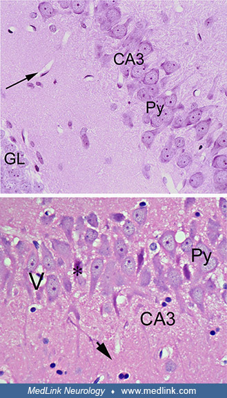
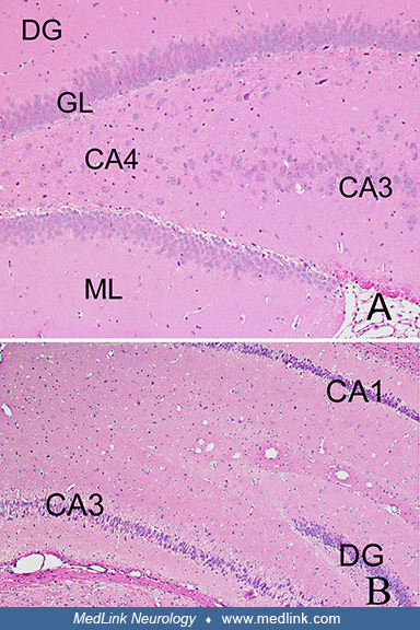
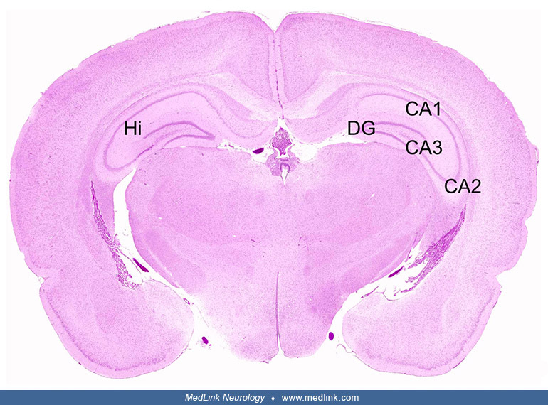
Cross-section of both hippocampal formations (Hi) showing the dentate gyrus (DG), and the CA3, CA2, and CA1 regions. Hematoxylin and eosin staining. (Source: Pulido 2008. Creative Commons Attribution license. http://creativecommons.org/licenses/by/3.0.)