Neuroimmunology
Leptospirosis
May. 10, 2025
MedLink, LLC
3525 Del Mar Heights Rd, Ste 304
San Diego, CA 92130-2122
Toll Free (U.S. + Canada): 800-452-2400
US Number: +1-619-640-4660
Support: service@medlink.com
Editor: editor@medlink.com
ISSN: 2831-9125
Toll Free (U.S. + Canada): 800-452-2400
US Number: +1-619-640-4660
Support: service@medlink.com
Editor: editor@medlink.com
ISSN: 2831-9125
Nearly 3,000 illustrations, including video clips of neurologic disorders.
Every article is reviewed by our esteemed Editorial Board for accuracy and currency.
Full spectrum of neurology in 1,200 comprehensive articles.
Listen to MedLink on the go with Audio versions of each article.
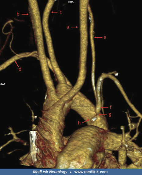
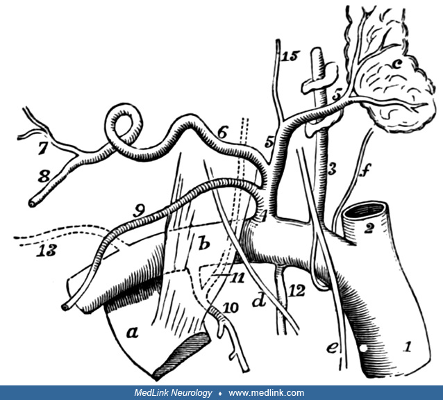
This diagram shows another variation in development of the aortic arch and great vessels (persistence of the distal portion of the right aortic arch, resulting in the origin of the right subclavian artery from the descending aorta, distal to the left subclavian origin): (A) aorta, (LS) left subclavian artery (P) pulmonary artery, (RS) right subclavian artery, (RV) right vertebral artery. Persistence of the distal portion of the right aortic arch and disappearance of a portion of its proximal part produces an apparent origin of the right subclavian artery from the descending aorta. (Contributed by Dr. Douglas Lanska. Source: Dwight T, McMurrich JP, Hamann CA, Piersol GA, White JW, Heisler JC. Human Anatomy: Including Structure and Development and Practical Considerations. Vol. 1. Philadelphia and London: J.B. Lippincott Co., 1907.)