General Neurology
Failed back surgery syndrome
Aug. 15, 2022
MedLink, LLC
3525 Del Mar Heights Rd, Ste 304
San Diego, CA 92130-2122
Toll Free (U.S. + Canada): 800-452-2400
US Number: +1-619-640-4660
Support: service@medlink.com
Editor: editor@medlink.com
ISSN: 2831-9125
Toll Free (U.S. + Canada): 800-452-2400
US Number: +1-619-640-4660
Support: service@medlink.com
Editor: editor@medlink.com
ISSN: 2831-9125
Nearly 3,000 illustrations, including video clips of neurologic disorders.
Every article is reviewed by our esteemed Editorial Board for accuracy and currency.
Full spectrum of neurology in 1,200 comprehensive articles.
Listen to MedLink on the go with Audio versions of each article.
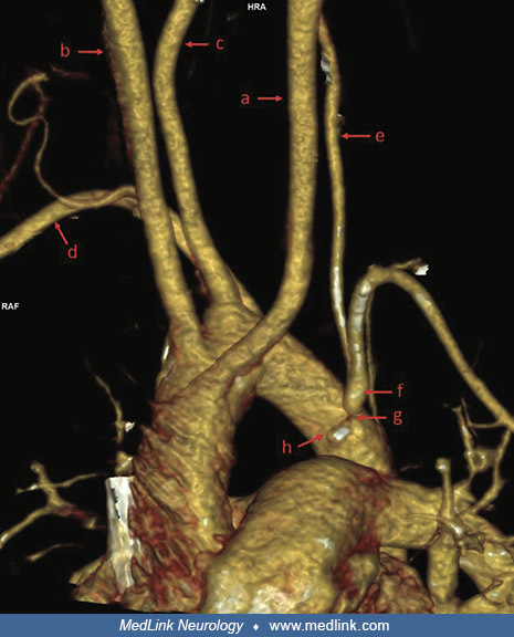
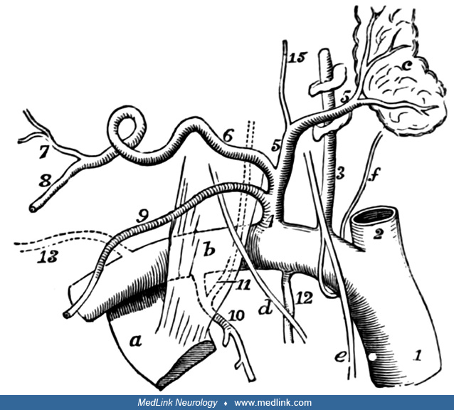
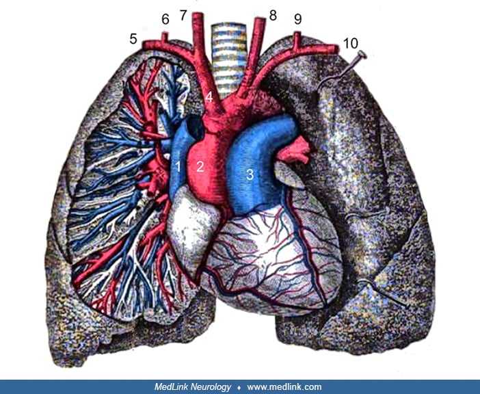
This diagram shows the aortic arch with a standard branching pattern of the great vessels, including the innominate (brachiocephalic) artery, left common carotid artery, and the left subclavian artery. The innominate artery branches into the right subclavian artery and the right common carotid artery. The first branch of the subclavian artery is the vertebral artery, which ascends behind the common carotid artery on both sides. (Contributed by Dr. Douglas Lanska. Source: Buchanon AM. Manual of Anatomy: Systematic and Practical, Including Embryology. Third edition. Volume 2. St. Louis: C.V. Mosby, 1917.)