Sleep Disorders
Sleep and headaches
Aug. 17, 2025
MedLink, LLC
3525 Del Mar Heights Rd, Ste 304
San Diego, CA 92130-2122
Toll Free (U.S. + Canada): 800-452-2400
US Number: +1-619-640-4660
Support: service@medlink.com
Editor: editor@medlink.com
ISSN: 2831-9125
Toll Free (U.S. + Canada): 800-452-2400
US Number: +1-619-640-4660
Support: service@medlink.com
Editor: editor@medlink.com
ISSN: 2831-9125
Nearly 3,000 illustrations, including video clips of neurologic disorders.
Every article is reviewed by our esteemed Editorial Board for accuracy and currency.
Full spectrum of neurology in 1,200 comprehensive articles.
Listen to MedLink on the go with Audio versions of each article.
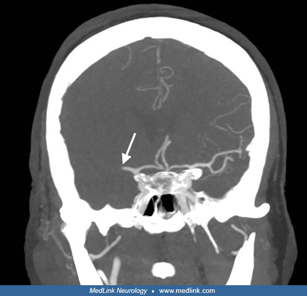
After injection of the mixture of iodine-based contrast agent, local anesthetic, and glucocorticoid, a final axial CT shows the contrast agent around the nerve sheath (white arrow) as well as in the epidural space (black arrows). The distribution of the local anesthetic and the glucocorticoid can be assumed to be identical to the contrast agent. (Contributed by Kristine Ann Blackham MD.)