Neuroimmunology
GAD antibody-spectrum disorders
Feb. 29, 2024
MedLink®, LLC
3525 Del Mar Heights Rd, Ste 304
San Diego, CA 92130-2122
Toll Free (U.S. + Canada): 800-452-2400
US Number: +1-619-640-4660
Support: service@medlink.com
Editor: editor@medlink.com
ISSN: 2831-9125
Toll Free (U.S. + Canada): 800-452-2400
US Number: +1-619-640-4660
Support: service@medlink.com
Editor: editor@medlink.com
ISSN: 2831-9125
Nearly 3,000 illustrations, including video clips of neurologic disorders.
Every article is reviewed by our esteemed Editorial Board for accuracy and currency.
Full spectrum of neurology in 1,200 comprehensive articles.
Listen to MedLink on the go with Audio versions of each article.
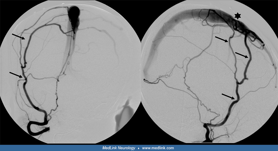
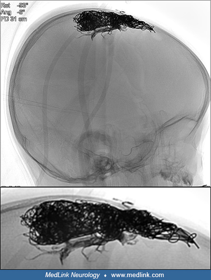
Following transvenous coiling of the superior sagittal sinus and distal liquid embolic embolization via the middle meningeal artery, the lateral unsubtracted view of the skull shows the large radiopaque (black) coil mass in the superior sagittal sinus at the level of the fistula point. The liquid embolic material is also black and is dispersed within the coil interstices. (Contributed by Kristine Ann Blackham MD.)