Neuroimmunology
CLIPPERS
Dec. 08, 2023
MedLink®, LLC
3525 Del Mar Heights Rd, Ste 304
San Diego, CA 92130-2122
Toll Free (U.S. + Canada): 800-452-2400
US Number: +1-619-640-4660
Support: service@medlink.com
Editor: editor@medlink.com
ISSN: 2831-9125
Toll Free (U.S. + Canada): 800-452-2400
US Number: +1-619-640-4660
Support: service@medlink.com
Editor: editor@medlink.com
ISSN: 2831-9125
Nearly 3,000 illustrations, including video clips of neurologic disorders.
Every article is reviewed by our esteemed Editorial Board for accuracy and currency.
Full spectrum of neurology in 1,200 comprehensive articles.
Listen to MedLink on the go with Audio versions of each article.
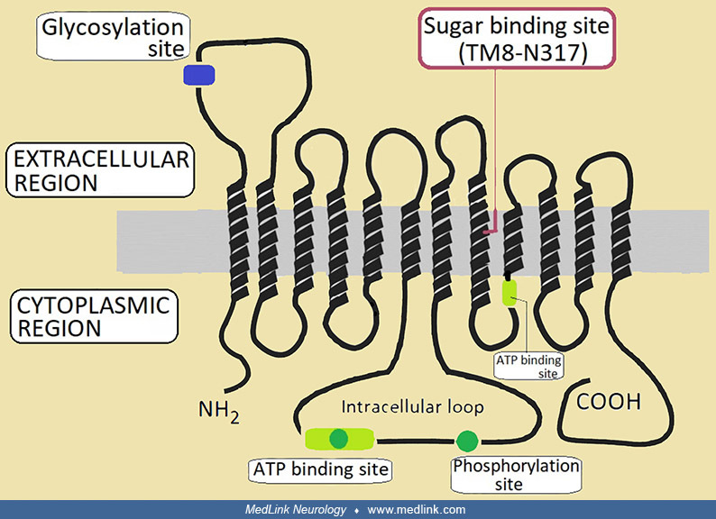
Schematic diagram of the GLUT1 protein structure, showing locations of specific mutations with corresponding phenotypes. The GLUT1 protein structure consists of 12 transmembrane domains and intracellular amino- and carboxy-termini (Mueckler et al 1985). Known GLUT1 deficiency syndrome (Klepper and Leiendecker 2007) and paroxysmal exercise-induced dyskinesia/epilepsy mutations are marked on this figure (yellow colored circles). The PED/epilepsy mutations from the pedigrees in Figure 2 are boxed. The solid bars indicate the locations of the intron-exon boundaries in the GLUT1 gene. Splice-site mutations are indicated at these solid bars with an asterisk. (Source: Suls et al 2008. Courtesy of Dr. Wim Van Paesschen, Department of Neurology, University Hospital Gasthuisberg, Katholieke Universiteit Leuven, Leuven, Belgium. Used with permission.)