Neuro-Oncology
Vestibular schwannoma
May. 27, 2026
MedLink, LLC
3525 Del Mar Heights Rd, Ste 304
San Diego, CA 92130-2122
Toll Free (U.S. + Canada): 800-452-2400
US Number: +1-619-640-4660
Support: service@medlink.com
Editor: editor@medlink.com
ISSN: 2831-9125
Toll Free (U.S. + Canada): 800-452-2400
US Number: +1-619-640-4660
Support: service@medlink.com
Editor: editor@medlink.com
ISSN: 2831-9125
Nearly 3,000 illustrations, including video clips of neurologic disorders.
Every article is reviewed by our esteemed Editorial Board for accuracy and currency.
Full spectrum of neurology in 1,200 comprehensive articles.
Listen to MedLink on the go with Audio versions of each article.
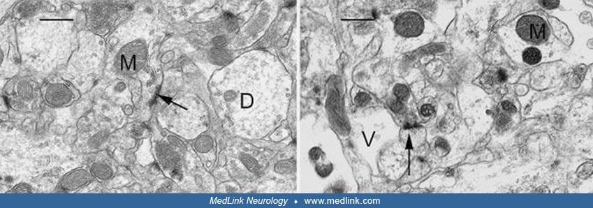
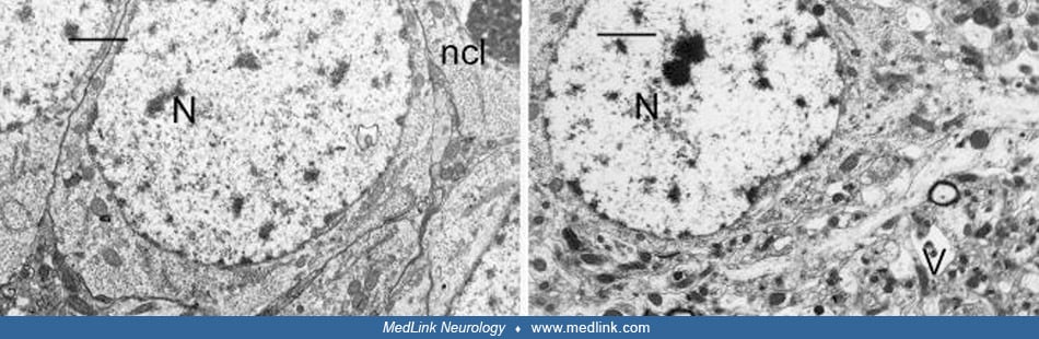
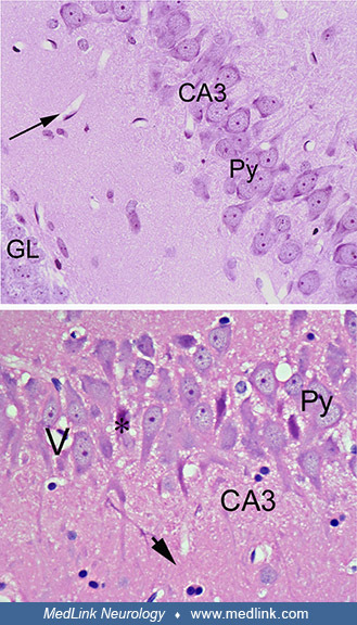
(Left) High magnification of the CA3 region of the hippocampus of a control rat showing good preservation and structural integrity. An electrodense dendritic spine (arrow), terminal axon (At), dendrite, and mitochondria (M) are identified. Scale Bar = x 0.6μm. (Right) High magnification of the CA3 region of the hippocampus of a rat treated by gavage with domoic acid for 64 days, showing loss of structural integrity and marked vacuolar (V) dilatation of dendrites. Scale Bar = x 0.6μm. (Source: Pulido 2008. Creative Commons Attribution license. http://creativecommons.org/licenses/by/3.0.)