Headache & Pain
Headache attributed to head trauma
Dec. 17, 2025
MedLink, LLC
3525 Del Mar Heights Rd, Ste 304
San Diego, CA 92130-2122
Toll Free (U.S. + Canada): 800-452-2400
US Number: +1-619-640-4660
Support: service@medlink.com
Editor: editor@medlink.com
ISSN: 2831-9125
Toll Free (U.S. + Canada): 800-452-2400
US Number: +1-619-640-4660
Support: service@medlink.com
Editor: editor@medlink.com
ISSN: 2831-9125
Nearly 3,000 illustrations, including video clips of neurologic disorders.
Every article is reviewed by our esteemed Editorial Board for accuracy and currency.
Full spectrum of neurology in 1,200 comprehensive articles.
Listen to MedLink on the go with Audio versions of each article.
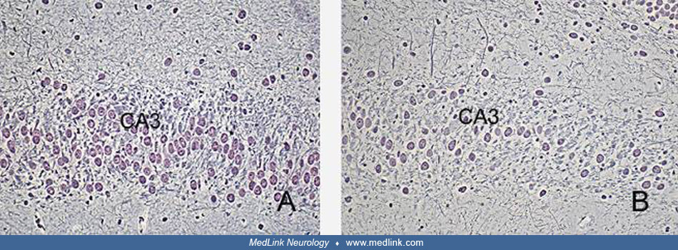
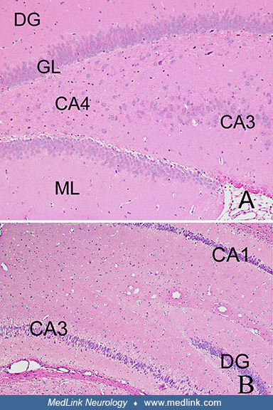
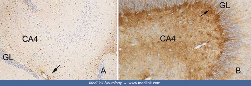
Brains of monkeys (Macaca fascicularis, the crab-eating macaque). Histological sections were processed for GFAP immunohistochemistry. (Left) Hippocampus of a control monkey showing the granular cell layer (GL) of the dentate gyrus and the CA4 region with spaced astrocytes labeled by glial fibrillary acidic protein (GFAP) immunohistochemistry. Some immunolabeled astrocytes are clearly identified around blood vessels (arrow). Objective x10. (Right) Hippocampus of a monkey treated with a single intravenous dose of domoic acid. The initial symptoms of toxicity lasted 90 minutes and included vomiting, gagging, lethargy, and disorientation. Necropsy was conducted 6 months after the injection. Sections show marked astrocytosis as revealed by the intensity of the GFAP immunohistochemical staining seen in CA4 and the subgranular zone (arrow); granular cell layer (GL). Objective x20. (Source: Pulido 2008. Creative Commons Attribution license. http://creativecommons.org/licenses/by/3.0.)