General Child Neurology
Kawasaki disease
Jan. 25, 2025
MedLink, LLC
3525 Del Mar Heights Rd, Ste 304
San Diego, CA 92130-2122
Toll Free (U.S. + Canada): 800-452-2400
US Number: +1-619-640-4660
Support: service@medlink.com
Editor: editor@medlink.com
ISSN: 2831-9125
Toll Free (U.S. + Canada): 800-452-2400
US Number: +1-619-640-4660
Support: service@medlink.com
Editor: editor@medlink.com
ISSN: 2831-9125
Nearly 3,000 illustrations, including video clips of neurologic disorders.
Every article is reviewed by our esteemed Editorial Board for accuracy and currency.
Full spectrum of neurology in 1,200 comprehensive articles.
Listen to MedLink on the go with Audio versions of each article.
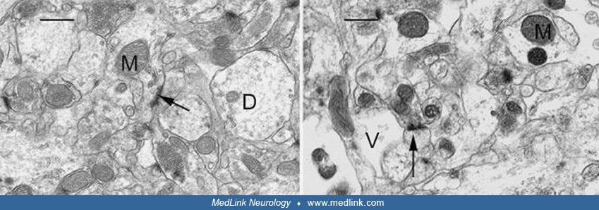
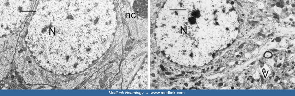
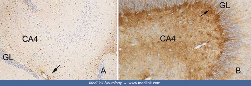
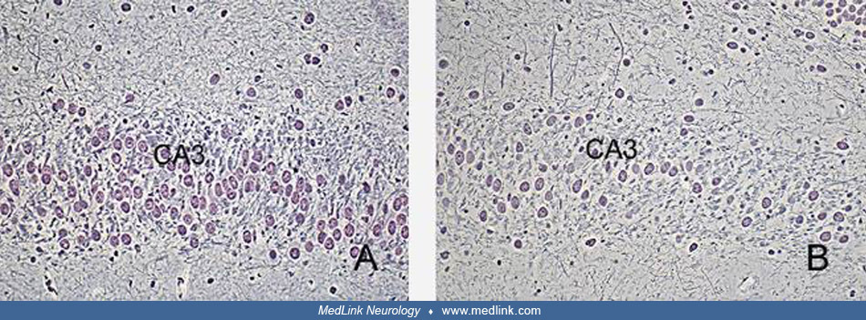
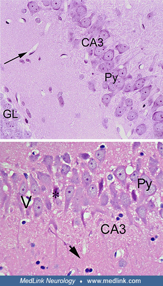
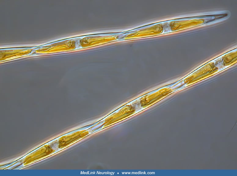
Brains of monkeys (Macaca fascicularis, the crab-eating macaque). (Left) Section of the CA3 region of the hippocampus of a control monkey, showing well-preserved pyramidal cells (Py). Blood vessels are seen as white spaces with the endothelial cells at the periphery (arrow), Hematoxylin and eosin staining. Objective x40. (Right) Sections of the hippocampus of a monkey treated with domoic acid, showing cell dropout and neuronal necrosis. Most pyramidal neurons appear with vacuolar cytoplasm (V). Some nuclear pyknosis (arrowhead) is also present. Hematoxylin and eosin staining. Objective x40. (Source: Pulido 2008. Creative Commons Attribution license (http://creativecommons.org/licenses/by/3.0.)