Neuropharmacology & Neurotherapeutics
Methylphenidate
Jun. 17, 2021
MedLink, LLC
3525 Del Mar Heights Rd, Ste 304
San Diego, CA 92130-2122
Toll Free (U.S. + Canada): 800-452-2400
US Number: +1-619-640-4660
Support: service@medlink.com
Editor: editor@medlink.com
ISSN: 2831-9125
Toll Free (U.S. + Canada): 800-452-2400
US Number: +1-619-640-4660
Support: service@medlink.com
Editor: editor@medlink.com
ISSN: 2831-9125
Nearly 3,000 illustrations, including video clips of neurologic disorders.
Every article is reviewed by our esteemed Editorial Board for accuracy and currency.
Full spectrum of neurology in 1,200 comprehensive articles.
Listen to MedLink on the go with Audio versions of each article.
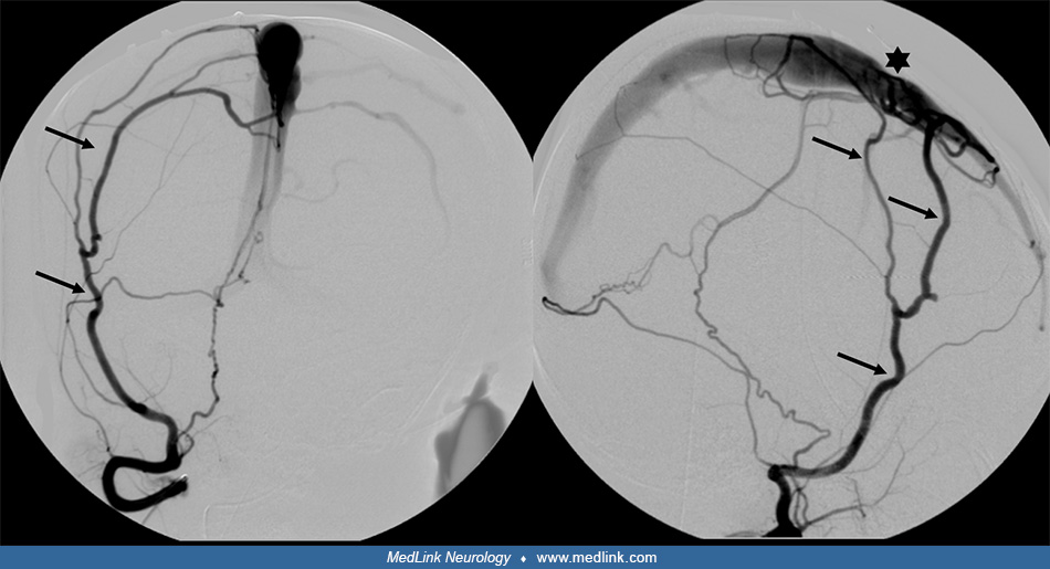
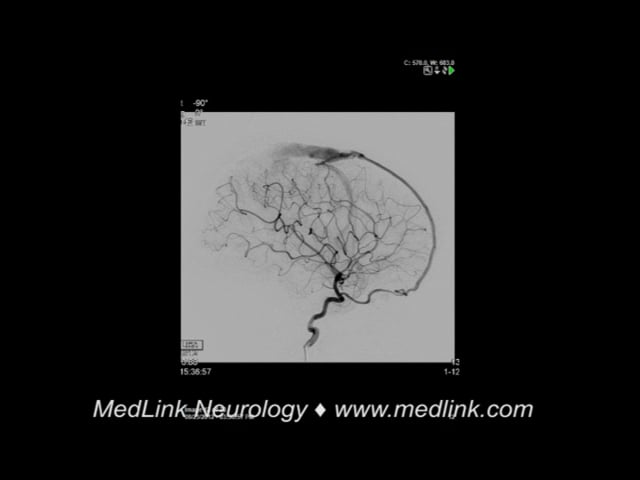
Anteroposterior (AP) and lateral views of the right external carotid artery injection show a hypertrophied middle meningeal artery (AP image 10 of 21, 1 sec). The enlarged middle meningeal artery has a fistulous connection with the superior sagittal sinus, which results in early and abnormal contrast opacification of the superior sagittal sinus (AP image 12 of 21, 1 sec, and lateral image 13 of 21, 7 sec). The fistulous drainage continues into the right transverse and sigmoid sinuses and right internal jugular vein (AP image 21 of 21, 3 sec). This “arterialization” of the sinuses renders them incapable of receiving the normal venous drainage of the brain. (Contributed by Dr. Kristine Ann Blackham.)