General Neurology
Neuropsychiatric symptoms associated with neurologic diseases
Feb. 13, 2026
MedLink, LLC
3525 Del Mar Heights Rd, Ste 304
San Diego, CA 92130-2122
Toll Free (U.S. + Canada): 800-452-2400
US Number: +1-619-640-4660
Support: service@medlink.com
Editor: editor@medlink.com
ISSN: 2831-9125
Toll Free (U.S. + Canada): 800-452-2400
US Number: +1-619-640-4660
Support: service@medlink.com
Editor: editor@medlink.com
ISSN: 2831-9125
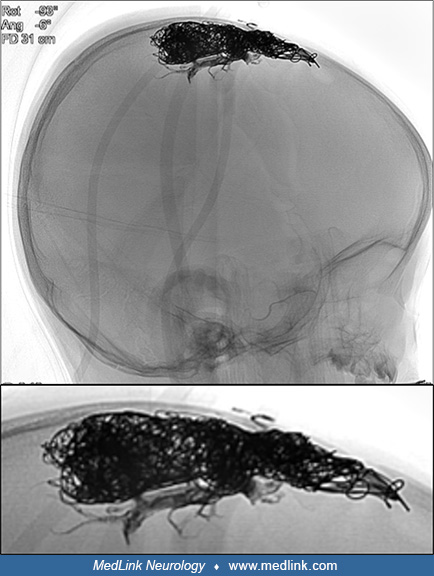
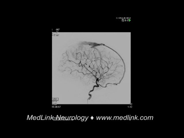
Nearly 3,000 illustrations, including video clips of neurologic disorders.
Every article is reviewed by our esteemed Editorial Board for accuracy and currency.
Full spectrum of neurology in 1,200 comprehensive articles.
Listen to MedLink on the go with Audio versions of each article.
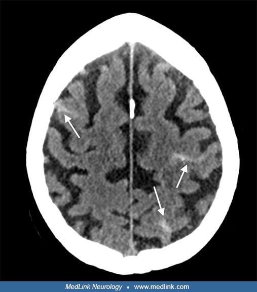
Axial MRI with diffusion-weighted images (DWI; b=1000 images are displayed) shows a punctate focus of diffusion restriction in the right parietal lobe (left image, dashed arrow) and small areas of diffusion restriction in the right basal ganglia (right image, straight arrows). Signal reduction in the corresponding apparent diffusion coefficient map (ADC; not displayed) confirmed these to be areas of acute infarction. However, the majority of the right middle cerebral artery territory is unaffected. (Contributed by Kristine Ann Blackham MD.)