Neuroimmunology
Multiple sclerosis: neuroimmunology
Nov. 15, 2025
MedLink, LLC
3525 Del Mar Heights Rd, Ste 304
San Diego, CA 92130-2122
Toll Free (U.S. + Canada): 800-452-2400
US Number: +1-619-640-4660
Support: service@medlink.com
Editor: editor@medlink.com
ISSN: 2831-9125
Toll Free (U.S. + Canada): 800-452-2400
US Number: +1-619-640-4660
Support: service@medlink.com
Editor: editor@medlink.com
ISSN: 2831-9125
Nearly 3,000 illustrations, including video clips of neurologic disorders.
Every article is reviewed by our esteemed Editorial Board for accuracy and currency.
Full spectrum of neurology in 1,200 comprehensive articles.
Listen to MedLink on the go with Audio versions of each article.
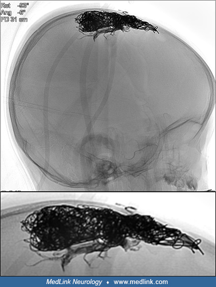
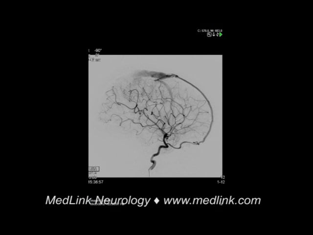
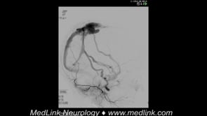
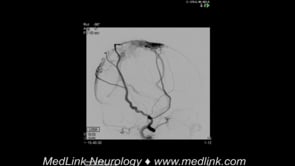
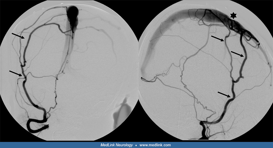
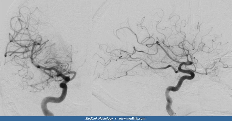
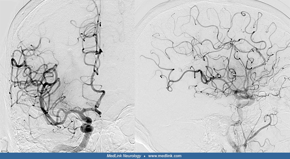
Left image: a lateral digital subtraction angiography image in the arterial phase of contrast injection in the right common carotid artery shows ectasia of the anterior falcine artery arising from the ophthalmic artery (arrow), which gives rise to a high-flow arteriovenous shunt as it connects directly to the superior sagittal sinus (arrowhead). Early venous drainage, which is opacification of the veins during the arterial phase of contrast administration, is the hallmark of an arteriovenous shunt. Right image: post-procedure, the shunt is gone, and there is no early venous drainage. (Contributed by Kristine Ann Blackham MD.)