Neuromuscular Disorders
Chronic progressive external ophthalmoplegia
Nov. 03, 2025
MedLink, LLC
3525 Del Mar Heights Rd, Ste 304
San Diego, CA 92130-2122
Toll Free (U.S. + Canada): 800-452-2400
US Number: +1-619-640-4660
Support: service@medlink.com
Editor: editor@medlink.com
ISSN: 2831-9125
Toll Free (U.S. + Canada): 800-452-2400
US Number: +1-619-640-4660
Support: service@medlink.com
Editor: editor@medlink.com
ISSN: 2831-9125
Nearly 3,000 illustrations, including video clips of neurologic disorders.
Every article is reviewed by our esteemed Editorial Board for accuracy and currency.
Full spectrum of neurology in 1,200 comprehensive articles.
Listen to MedLink on the go with Audio versions of each article.
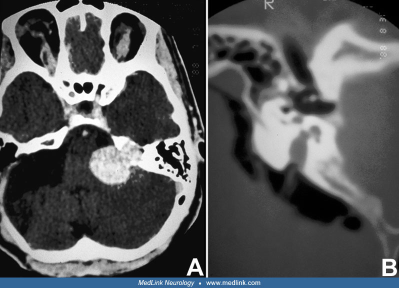
(A) Axial, enhanced image at the level of the cerebellopontine angle, demonstrating a large, diffusely enhancing tumor centered over the internal auditory canal. The mass extends through the porus acusticus into the internal enlarged auditory canal. Note the compression of the nearby pons and cerebellum. There is little, if any, associated edema within the brain, suggesting that compression developed slowly. (B) Axial image using bone windows and air contrast, demonstrating a small, intracanalicular tumor within the internal auditory canal. (Contributed by Dr. Herbert Newton.)