Neuromuscular Disorders
Emery-Dreifuss muscular dystrophy
Sep. 25, 2025
MedLink, LLC
3525 Del Mar Heights Rd, Ste 304
San Diego, CA 92130-2122
Toll Free (U.S. + Canada): 800-452-2400
US Number: +1-619-640-4660
Support: service@medlink.com
Editor: editor@medlink.com
ISSN: 2831-9125
Toll Free (U.S. + Canada): 800-452-2400
US Number: +1-619-640-4660
Support: service@medlink.com
Editor: editor@medlink.com
ISSN: 2831-9125
Nearly 3,000 illustrations, including video clips of neurologic disorders.
Every article is reviewed by our esteemed Editorial Board for accuracy and currency.
Full spectrum of neurology in 1,200 comprehensive articles.
Listen to MedLink on the go with Audio versions of each article.
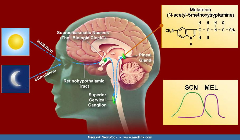
Melatonin (upper right inset) is produced in the pineal gland. The production and secretion of melatonin are mediated largely by postganglionic retinal nerve fibers that pass through the retinohypothalamic tract to the suprachiasmatic nucleus, then to the superior cervical ganglion, and finally to the pineal gland. This neuronal system is activated by darkness and suppressed by light (left insets). The activation of alpha1 and beta1 adrenergic receptors in the pineal gland raises cyclic AMP, leading to the release of melatonin. The daily rhythm of melatonin secretion is controlled by the endogenous master pacemaker located in the suprachiasmatic nuclei. The lower right inset shows the temporal relationship between the activity of the suprachiasmatic nuclei and the secretion of melatonin within a period of 24 hours (not to scale). SCN – suprachiasmatic nuclei, MEL – melatonin. (Adapted 2006, with permission, from: Brzezinski A. Mechanisms of disease: melatonin in humans. New Engl J Med 1997;336:186-195. Copyright © 1997 Massachusetts Medical Society.)