Neuro-Ophthalmology & Neuro-Otology
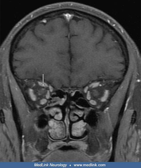
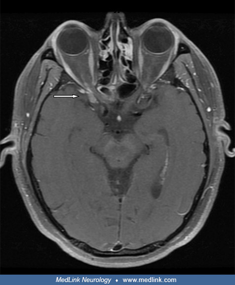
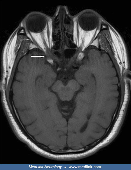
Vogt-Koyanagi-Harada syndrome
Jun. 27, 2025
MedLink, LLC
3525 Del Mar Heights Rd, Ste 304
San Diego, CA 92130-2122
Toll Free (U.S. + Canada): 800-452-2400
US Number: +1-619-640-4660
Support: service@medlink.com
Editor: editor@medlink.com
ISSN: 2831-9125
Toll Free (U.S. + Canada): 800-452-2400
US Number: +1-619-640-4660
Support: service@medlink.com
Editor: editor@medlink.com
ISSN: 2831-9125
Nearly 3,000 illustrations, including video clips of neurologic disorders.
Every article is reviewed by our esteemed Editorial Board for accuracy and currency.
Full spectrum of neurology in 1,200 comprehensive articles.
Listen to MedLink on the go with Audio versions of each article.
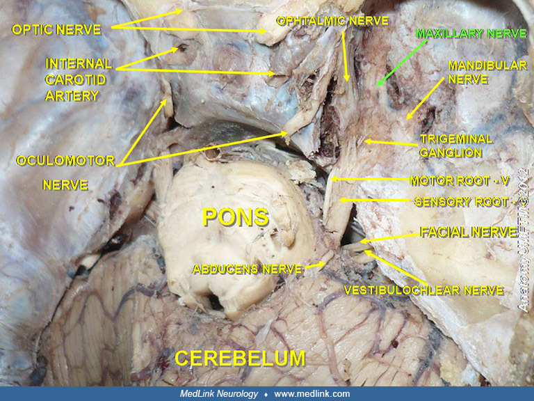
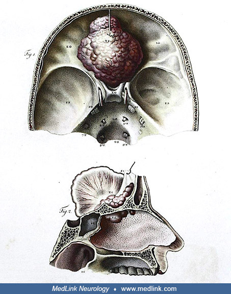
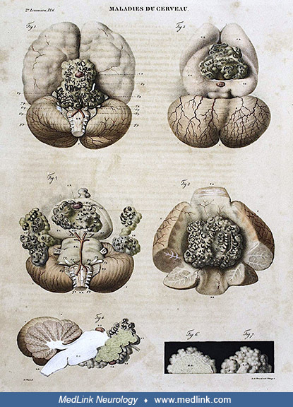
Lithograph plate from Cruveilhier’s “Anatomie Pathologique du Corps Humain” shows pearled tumors of the skull base (epidermoid tumors) involving all the cranial nerves and the pituitary gland and stalk (Cruveilhier 1829-1853). (Image edited by Dr. Douglas J Lanska to improve exposure and contrast, and to correct a color cast. Public domain.)