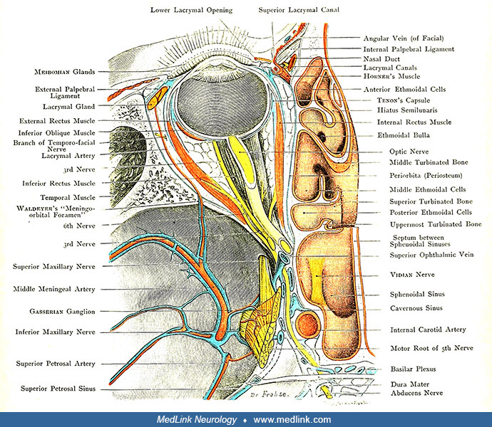
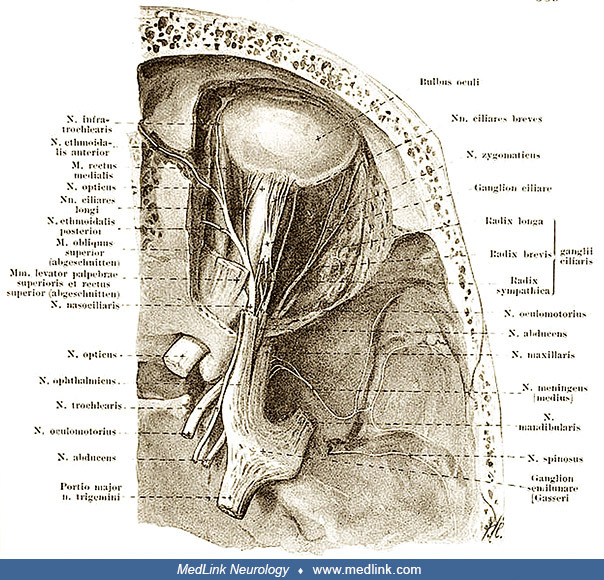
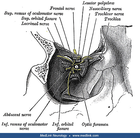
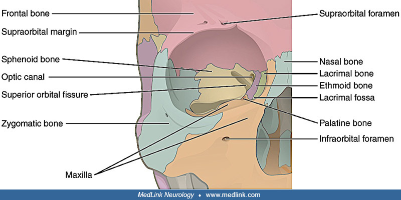
General Child Neurology
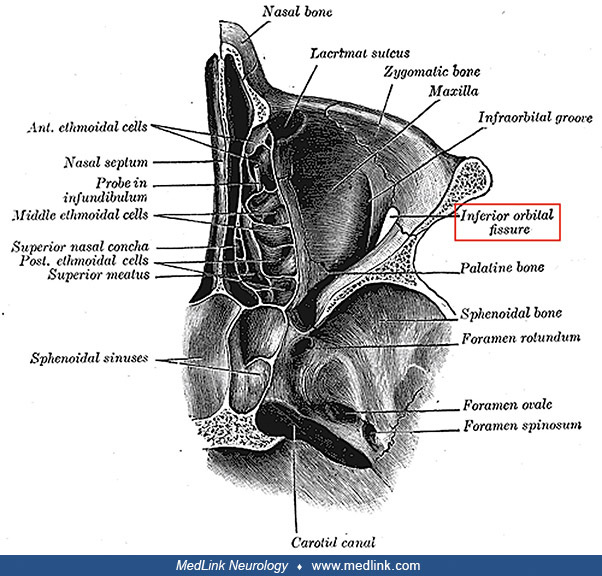
Fetal alcohol syndrome
Jan. 21, 2026
MedLink, LLC
3525 Del Mar Heights Rd, Ste 304
San Diego, CA 92130-2122
Toll Free (U.S. + Canada): 800-452-2400
US Number: +1-619-640-4660
Support: service@medlink.com
Editor: editor@medlink.com
ISSN: 2831-9125
Toll Free (U.S. + Canada): 800-452-2400
US Number: +1-619-640-4660
Support: service@medlink.com
Editor: editor@medlink.com
ISSN: 2831-9125
Nearly 3,000 illustrations, including video clips of neurologic disorders.
Every article is reviewed by our esteemed Editorial Board for accuracy and currency.
Full spectrum of neurology in 1,200 comprehensive articles.
Listen to MedLink on the go with Audio versions of each article.
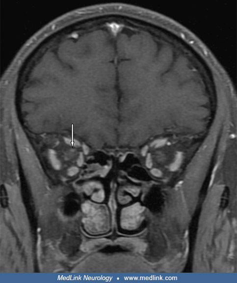
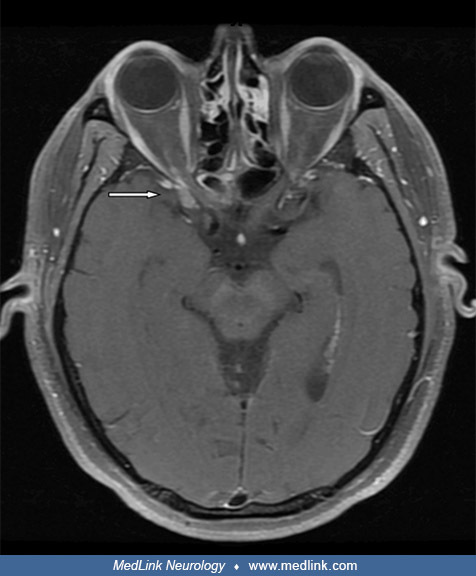
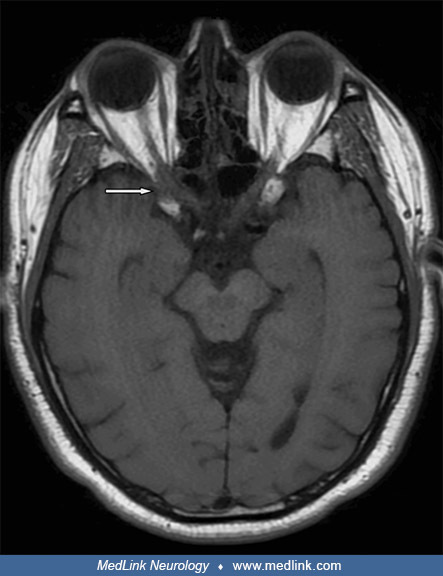
(A) An axial T2-weighted orbital MR image in a 67-year-old man misdiagnosed with Tolosa-Hunt syndrome showing mucosal thickening in the sphenoid sinus and hypointense areas in the center (yellow asterisk). (B, C) Orbital MR sections in axial T2-weighted and post-contrast fat-suppressed T1-weighted imaging reveal inflammatory signal alterations in the extraconal adipose tissue (yellow arrow), along with thickening and enhanced contrast uptake in the extraocular muscles (red arrows) and optic nerve (white arrow). Extraconal indicates a location outside of the orbit's muscle cone, containing the eyeball, optic nerve, and major vessels, but still within the bony orbit, containing structures like the lacrimal gland, extraocular muscles, and fat. Additionally, there is contrast enhancement observed in the left cavernous sinus (red asterisk), suggesting Tolosa-Hunt syndrome or orbital pseudotumor in the left orbit. (Source: Karakeçili F, Barkay O, Sümer B, et al. Invasive aspergillosis with cavernous sinus thrombosis following high-dose corticosteroid therapy: a challenging case of rhino-orbital-cerebral mycosis. J Fungi [Basel] 2024;10[11]:788. Creative Commons Attribution 4.0 International [CC BY 4.0] license, creativecommons.org/licenses/by/4.0.)