General Child Neurology
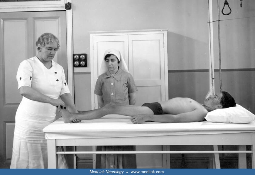
Neonatal white matter injury
Jan. 31, 2025
MedLink, LLC
3525 Del Mar Heights Rd, Ste 304
San Diego, CA 92130-2122
Toll Free (U.S. + Canada): 800-452-2400
US Number: +1-619-640-4660
Support: service@medlink.com
Editor: editor@medlink.com
ISSN: 2831-9125
Toll Free (U.S. + Canada): 800-452-2400
US Number: +1-619-640-4660
Support: service@medlink.com
Editor: editor@medlink.com
ISSN: 2831-9125
Nearly 3,000 illustrations, including video clips of neurologic disorders.
Every article is reviewed by our esteemed Editorial Board for accuracy and currency.
Full spectrum of neurology in 1,200 comprehensive articles.
Listen to MedLink on the go with Audio versions of each article.
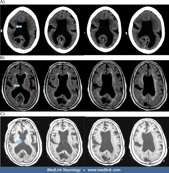
Brain imaging in an 85-year-old with schizencephaly with longstanding hemiparesis previously misdiagnosed as poliomyelitis. (A) CT brain without contrast showing inferior to superior sequence (shown left to right) of right-sided schizencephaly extending from the cortex to the right lateral ventricle. (B) MRI brain without contrast (axial T2 FLAIR) showing inferior to superior sequence (shown left to right) of right-sided schizencephaly extending from the cortex to the lateral ventricle and hypoplasia of the corpus callosum. (C) MRI brain without contrast (axial T1) showing margins of the schizencephaly defect lined with dysplastic gray matter. Arrows indicate a cleft extending from the right cerebral cortex into the lateral ventricle. (From: Guo Z, Scripko PD, Gertz AM. Open lip schizencephaly misdiagnosed as paralytic poliomyelitis in an 85-year-old: a case report. Clin Case Rep 2025;13[6]:e70556. Creative Commons Attribution 4.0 International [CC BY 4.0] license, creativecommons.org/licenses/by/4.0.)