Neuromuscular Disorders
Disorders of lipid metabolism
Sep. 25, 2025
MedLink, LLC
3525 Del Mar Heights Rd, Ste 304
San Diego, CA 92130-2122
Toll Free (U.S. + Canada): 800-452-2400
US Number: +1-619-640-4660
Support: service@medlink.com
Editor: editor@medlink.com
ISSN: 2831-9125
Toll Free (U.S. + Canada): 800-452-2400
US Number: +1-619-640-4660
Support: service@medlink.com
Editor: editor@medlink.com
ISSN: 2831-9125
Nearly 3,000 illustrations, including video clips of neurologic disorders.
Every article is reviewed by our esteemed Editorial Board for accuracy and currency.
Full spectrum of neurology in 1,200 comprehensive articles.
Listen to MedLink on the go with Audio versions of each article.
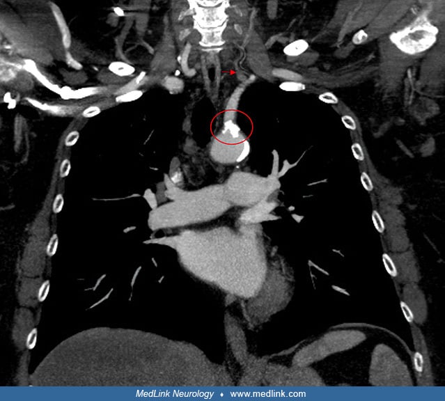
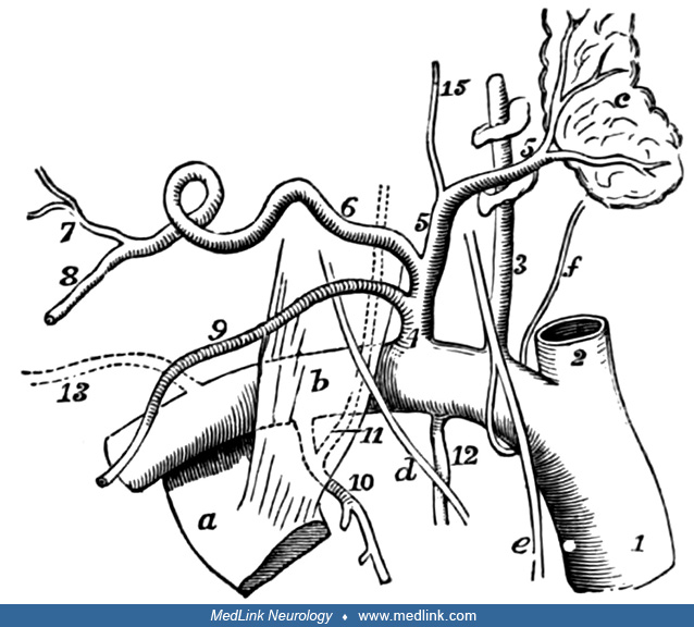
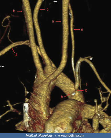
Angio-CT of the cephalad arteries in the spatial projection presentation from a 10-year-old girl with neurologic symptoms due to subclavian steal syndrome. The left common carotid artery (a), the right common carotid artery (b), the right vertebral artery (c), and the right subclavian artery (d) branch off from the aortic arch. The left subclavian artery (f), critically narrowed at the ostium (g), leaves in the immediate vicinity of the origin of the ductus arteriosus (h). The left vertebral body (e) is at least twice as narrow as the contralateral vertebral body (c). (Szmigielska A, Buczyński M, Śledziewska A, Małgorzata-Pańczyk-Tomaszewska, Furmanek MI. Transient ischaemic attacks in a girl with subclavian steal syndrome. J Mother Child 2025;29(1):101-5. Creative Commons Attribution 4.0 International [CC BY 4.0] license, creativecommons.org/licenses/by/4.0.)