Neuromuscular Disorders
Congenital myasthenic syndromes
Apr. 22, 2024
MedLink®, LLC
3525 Del Mar Heights Rd, Ste 304
San Diego, CA 92130-2122
Toll Free (U.S. + Canada): 800-452-2400
US Number: +1-619-640-4660
Support: service@medlink.com
Editor: editor@medlink.com
ISSN: 2831-9125
Toll Free (U.S. + Canada): 800-452-2400
US Number: +1-619-640-4660
Support: service@medlink.com
Editor: editor@medlink.com
ISSN: 2831-9125
Nearly 3,000 illustrations, including video clips of neurologic disorders.
Every article is reviewed by our esteemed Editorial Board for accuracy and currency.
Full spectrum of neurology in 1,200 comprehensive articles.
Listen to MedLink on the go with Audio versions of each article.
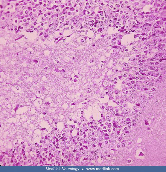
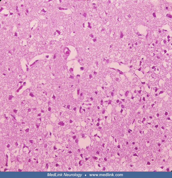
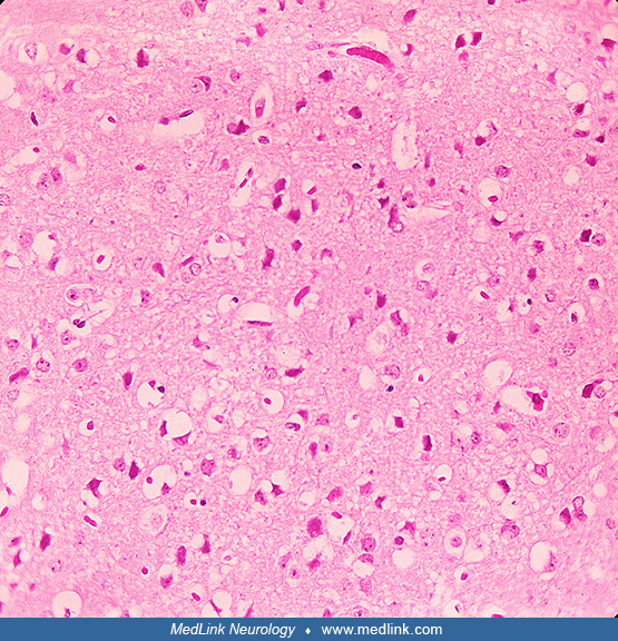
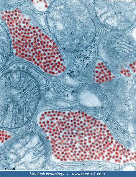
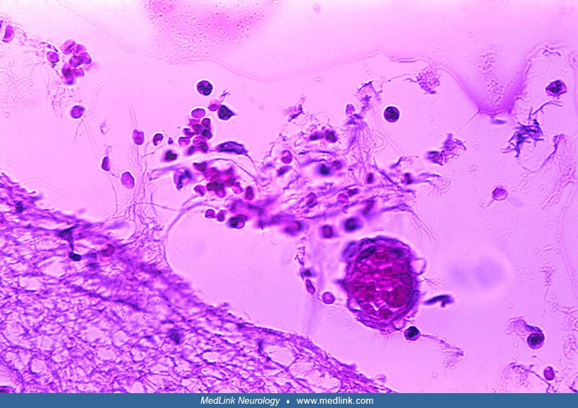
Histopathologic features from a 5-year-old girl with eastern equine encephalitis in 2005, as part of a study of eastern equine encephalitis in Massachusetts and New Hampshire from 1970 to 2010. The postmortem samples of central nervous system tissue were obtained 10 days after the onset of symptoms. Eastern equine encephalitis virus colocalizes with areas of tissue injury in the brain. Immunohistochemistry with eastern equine encephalitis immune ascites demonstrates eastern equine encephalitis viral antigens in the thalamus. Specificity for Eastern equine encephalitis virus immunoreactivity of this ascites fluid was confirmed by the lack of staining on control brain specimens. (Source: Silverman MA, Misasi J, Smole S, et al. Eastern equine encephalitis in children, Massachusetts and New Hampshire, USA, 1970-2010. Emerg Infect Dis 2013;19[2]:194-201. Public domain.)