General Neurology
Hemophilia and other coagulation disorders: neurologic aspects
Jan. 06, 2024
MedLink®, LLC
3525 Del Mar Heights Rd, Ste 304
San Diego, CA 92130-2122
Toll Free (U.S. + Canada): 800-452-2400
US Number: +1-619-640-4660
Support: service@medlink.com
Editor: editor@medlink.com
ISSN: 2831-9125
Toll Free (U.S. + Canada): 800-452-2400
US Number: +1-619-640-4660
Support: service@medlink.com
Editor: editor@medlink.com
ISSN: 2831-9125
Nearly 3,000 illustrations, including video clips of neurologic disorders.
Every article is reviewed by our esteemed Editorial Board for accuracy and currency.
Full spectrum of neurology in 1,200 comprehensive articles.
Listen to MedLink on the go with Audio versions of each article.
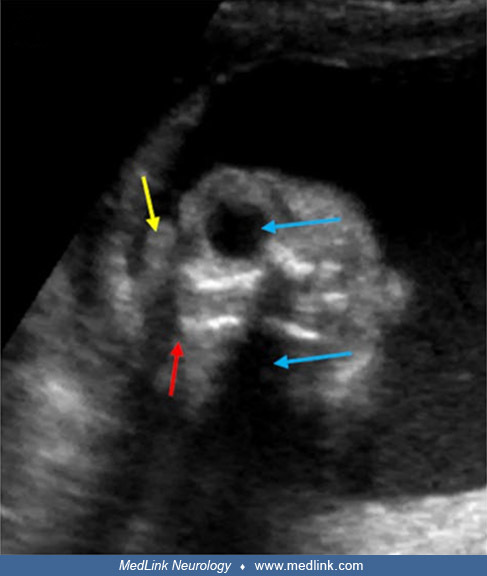
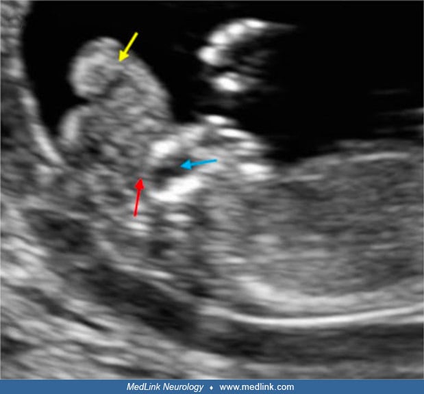
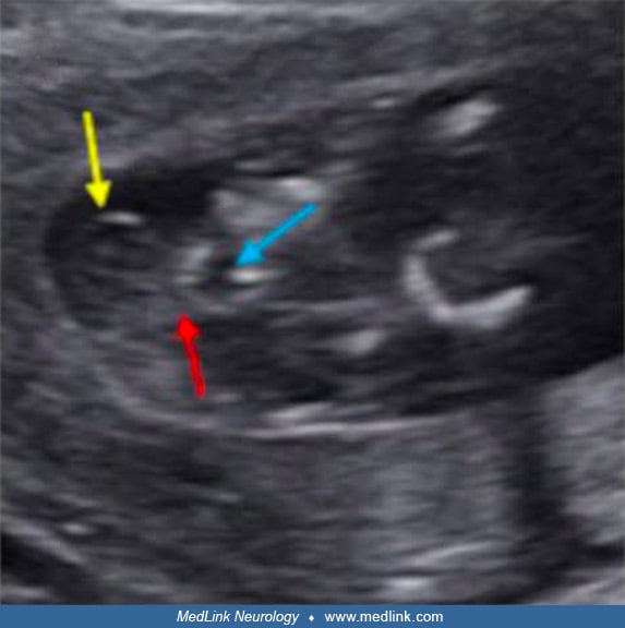
Two-dimensional ultrasound images at 17 weeks of gestation showing typical features of exencephaly (“Mickey Mouse” sign; white arrows). The calvarium is absent, and the brain is “floating” in the amniotic fluid. (Source: Sugiura T, Sato Y, Nakanami N, Tsukimori K. Prenatal sonographic image of sirenomelia with anencephaly and craniorachischisis totalis. Case Rep Obstet Gynecol 2018;2018:7058253. Creative Commons Attribution [CC BY 4.0] license, creativecommons.org/licenses/by/4.0.)