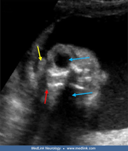
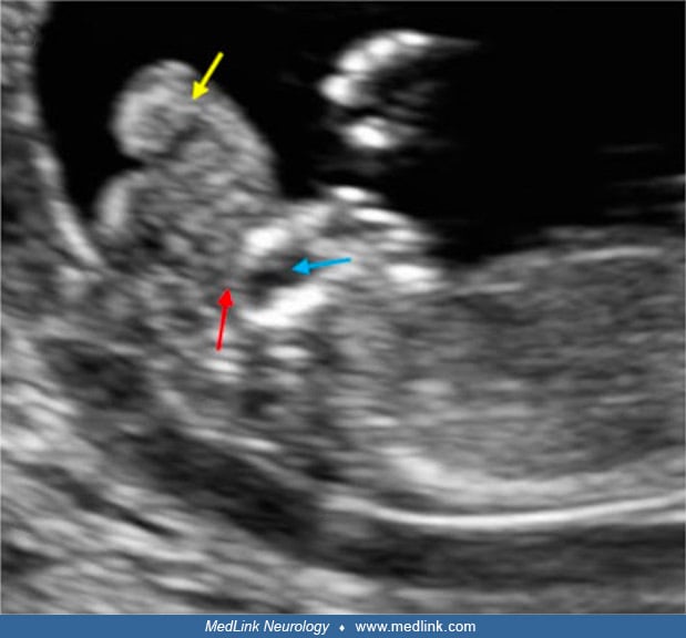
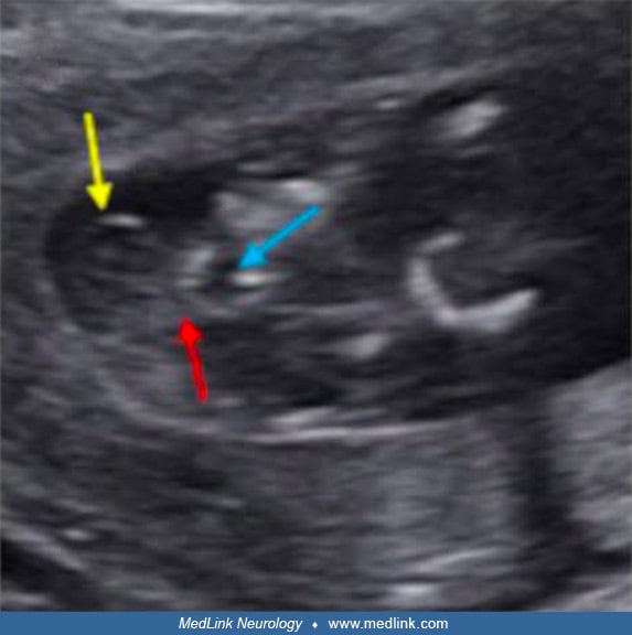
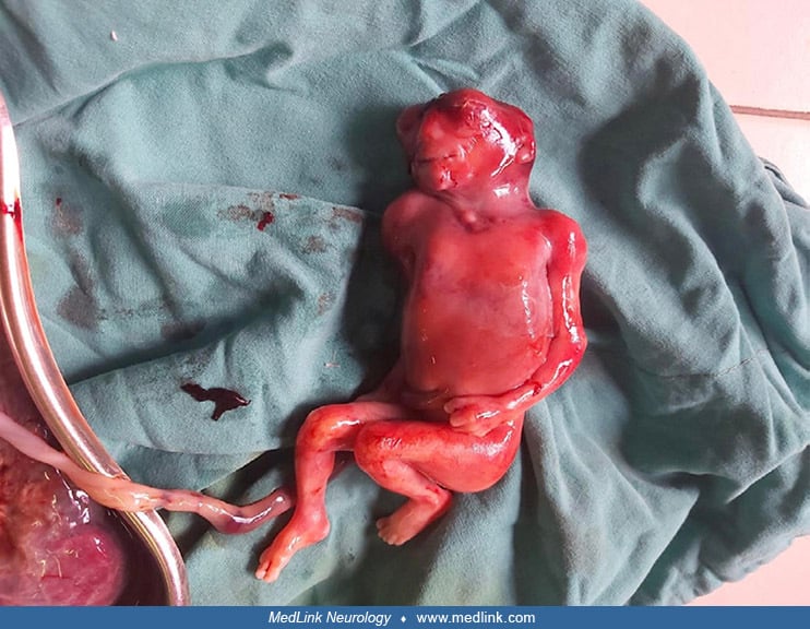
Headache & Pain
Pain
Aug. 14, 2024
MedLink®, LLC
3525 Del Mar Heights Rd, Ste 304
San Diego, CA 92130-2122
Toll Free (U.S. + Canada): 800-452-2400
US Number: +1-619-640-4660
Support: service@medlink.com
Editor: editor@medlink.com
ISSN: 2831-9125
Toll Free (U.S. + Canada): 800-452-2400
US Number: +1-619-640-4660
Support: service@medlink.com
Editor: editor@medlink.com
ISSN: 2831-9125
Nearly 3,000 illustrations, including video clips of neurologic disorders.
Every article is reviewed by our esteemed Editorial Board for accuracy and currency.
Full spectrum of neurology in 1,200 comprehensive articles.
Listen to MedLink on the go with Audio versions of each article.
Wall-eyed stereo view of ECG modeled into the folate-binding site of human dihydrofolate reductase (DHFR). Abbreviations: ECG: (-)-epicatechin gallate; DHFR: dihydrofolate reductase. Carbon atoms of the ECG ligand and surrounding protein are colored green and grey, respectively. Residue Phe-31, located behind the ECG, is unlabeled. Four different ligands from human and chicken DHFR crystal structures were used to define a binding envelope, shown in cyan. These were placed in a common orientation by superimposing backbone atoms from a common set of protein residues located around the ligands. (Source: Sanchez-del-Campo L, Sáez-Ayala M, Chazarra S, Cabezas-Herrera J, Rodríguez-López JN. Binding of natural and synthetic polyphenols to human dihydrofolate reductase. Int J Mol Sci 2009;10[12]:5398-410. Creative Commons Attribution [CC BY 3.0] license, creativecommons.org/licenses/by/3.0.)