Peripheral Neuropathies
Chronic idiopathic axonal polyneuropathy
Sep. 23, 2023
MedLink®, LLC
3525 Del Mar Heights Rd, Ste 304
San Diego, CA 92130-2122
Toll Free (U.S. + Canada): 800-452-2400
US Number: +1-619-640-4660
Support: service@medlink.com
Editor: editor@medlink.com
ISSN: 2831-9125
Toll Free (U.S. + Canada): 800-452-2400
US Number: +1-619-640-4660
Support: service@medlink.com
Editor: editor@medlink.com
ISSN: 2831-9125
Nearly 3,000 illustrations, including video clips of neurologic disorders.
Every article is reviewed by our esteemed Editorial Board for accuracy and currency.
Full spectrum of neurology in 1,200 comprehensive articles.
Listen to MedLink on the go with Audio versions of each article.
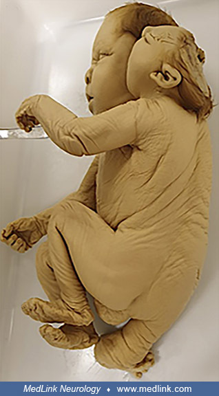
Normal (left) and exencephalic (right) fetal head in mouse model of congenital cranioschisis. Sagittal section of normal (left) and exencephalic (right) fetal head at embryonic day 17 (4x, hematoxylin and eosin). (Source: Oria M, Duru S, Figueira RL, et al. Cell necrosis, intrinsic apoptosis and senescence contribute to the progression of exencephaly to anencephaly in a mice model of congenital chranioschisis. Cell Death Dis 2019;10[10]:721. Creative Commons Attribution [CC BY 4.0] license, creativecommons.org/licenses/by/4.0.)